Article Text

Statistics from Altmetric.com
A 55-year-old woman, a known case of chronic rheumatic heart disease with mitral stenosis and atrial fibrillation with past history of closed mitral commisurotomy 18 years ago, was admitted with complaints of nausea, vomiting and dyspnoea. A 12-lead electrocardiogram revealed absence of ‘p’ waves, bradycardia and ‘inverted check mark’ sign with inverted ‘T’ wave not rising above baseline, suggesting digoxin toxicity which was confirmed with serum digoxin level (8 ng m/ml) (see supplementary ECG). Fluoroscopy during coronary angiography in posterior–anterior and lateral view revealed a rare ‘egg shell calcification’ of left atrial wall involving anterior mitral valve (figure 1; see supplementary video 1). Transthoracic and transesophageal echocardiography revealed mitral restenosis with mitral valve area of 0.8 cm2 and thickened and calcified interatrial septum (figure 2).

Left lateral fluoroscopic still image revealing a rare pattern of "egg shell calcification" of left atrium involving anterior mitral valve.
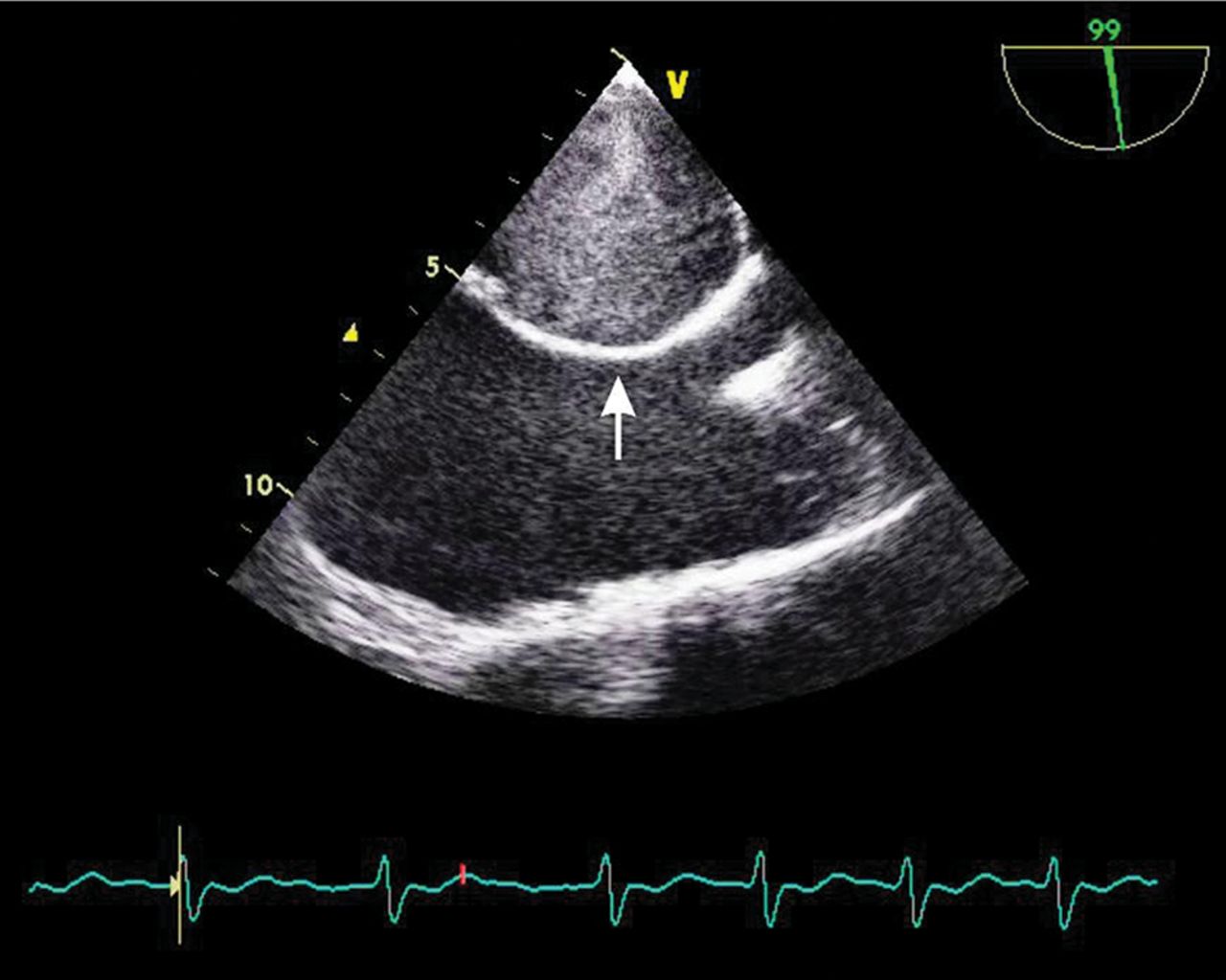
Transesophageal echocardiographic image revealing thickened and calcified interatrial septum (arrow).
This rare pattern of left atrium calcification predominantly affects women with chronic rheumatic mitral stenosis and atrial fibrillation of prolonged duration. Left atrial calcification has been classified as Type A (calcification confined to left atrial appendage), Type B (involves free wall of the left atrium and mitral valve), and Type C (small area of calcification confined to the posterior wall of the left atrium, known as McCallum's patch, resulting from a jet lesion because of mitral regurgitation).1
Complete calcification of left atrium has also been described as ‘coconut atrium’ or ‘porcelain atrium’, and should be anticipated before any intervention. Interatrial septal puncture during balloon mitral valvotomy is difficult due to thickened and calcified interatrial septum. Surgical difficulties include particle embolisation, difficult access to mitral valve and haemorrhage during closure of atrium.2
Supplementary materials
Supplementary Data
This web only file has been produced by the BMJ Publishing Group from an electronic file supplied by the author(s) and has not been edited for content.
Files in this Data Supplement:
- Data supplement 1 - Supplement ECG
- Data supplement 2 - Supplement Video
Footnotes
Contributors The first author has detected the finding and written the manuscript which was supported and edited by the second author.
Patient consent Obtained, consent from the patient’s relative was taken.
Competing interests None.
Provenance and peer review Not commissioned; internally peer reviewed.