Article Text

Statistics from Altmetric.com
A 48-year-old asymptomatic male, presented with mild elevation of transaminases and incidentally a basal oxygen saturation of 91% was registered. To rule out right to left shunting, the patient underwent a transoesophageal echocardiography. Initially, an intravenous cannula positioned on the left arm was used to inject agitated saline contrast. Unexpected opacification of the left atrium was observed (figure 1, online supplementary video 1). New injection into the right arm showed normal bubbles arrival into the right atrium without evidence of patent foramen ovale (online supplementary figure, video 2). Comprehensive Cardiac CT Angiography with intravenous cannula located in the right arm confirmed the presence of a persistent left superior vena cava draining into the left atrium (figure 2).1 ,2 The presence of left hemicranial and left upper limb venous drainage into the left atrium instead of the pulmonary circulation via the right heart chambers was the underlying cause of the oxygen desaturation. Transaminases levels reverted to normal values in the follow-up.

Transoesophageal echocardiography in mid-position view, 90° right; both atria are evident. Agitated saline contrast into the left arm resulted in opacification of the left atrium. LA, Left Atrium; RA, Right Atrium; AS, atrial septum; SVC, Superior Vena Cava.
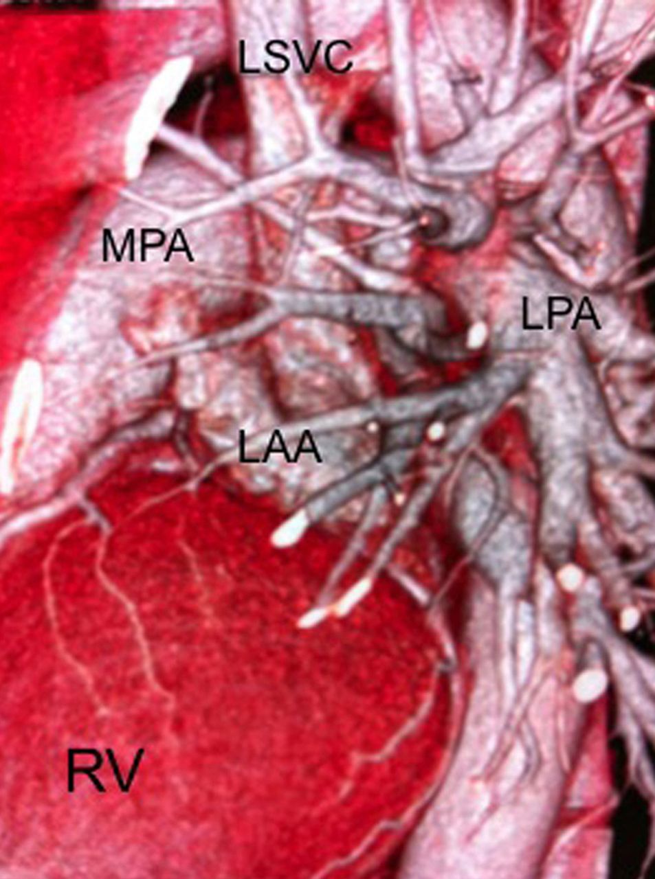
Cardiac CT Angiography, Three Dimensional Volume Rendering. Injection of Iodine-based contrast into the left arm was performed to have comprehensive information of cardiac vein drainage. Persistent Left Superior Vena Cava draining to the Left Atrium was demonstrated. LSVC, Left Superior Vena Cava; LAA, Left Atrial Appendage; RV, Right Ventricle; MPA, Main Pulmonary Artery; LPA, Left Pulmonary Artery.
References
Supplementary materials
Supplementary Data
This web only file has been produced by the BMJ Publishing Group from an electronic file supplied by the author(s) and has not been edited for content.
Files in this Data Supplement:
- Data supplement 1 - Online supplement
- Data supplement 2 - Online video 1
- Data supplement 3 - Online video 2
Footnotes
-
Contributors JGM had the original idea, selected and processed the images and videos and reviewed the manuscript; EGL wrote the manuscript; MAC and LA-P were responsible for the overall content as guarantors.
-
Funding None.
-
Competing interests None.
-
Patient consent Obtained.
-
Provenance and peer review Not commissioned; internally peer reviewed.