Article Text

Statistics from Altmetric.com
A 49-year-old man presented to our hospital with episodes of left-sided abdominal pain and fever. He had a medical history of past treated polycythaemia vera and old myocardial infarction. He had been treated with oral aspirin, and no other antithrombic agent was administrated. Physical examination was normal except for tenderness at the left upper quadrant of the abdomen. Chest x-ray and 12-lead ECG revealed no significant change. Laboratory data showed elevated C reactive protein (8.67 mg/dl) and D-dimer (6.4 μg/ml).
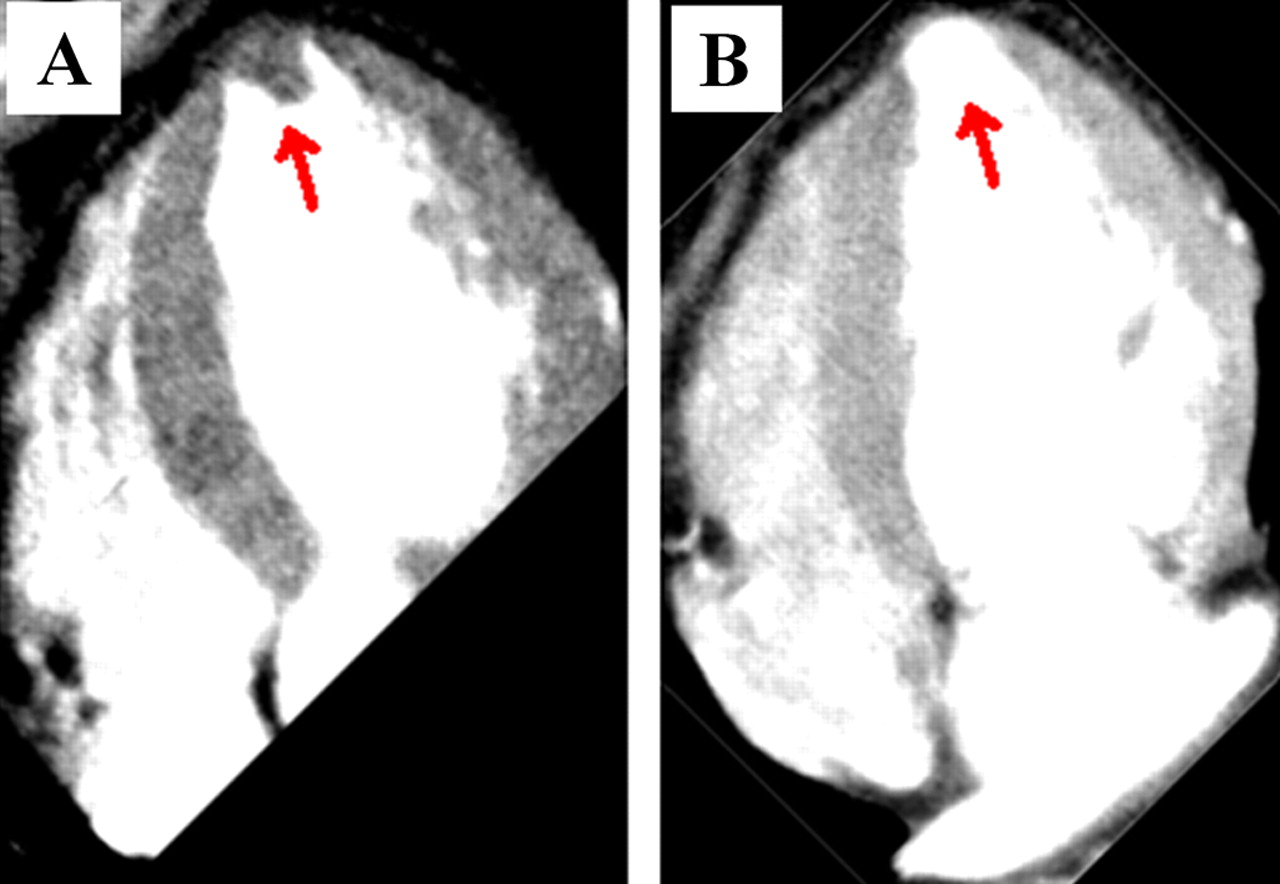
He immediately underwent contrast-enhanced CT, which revealed splenomegaly with broad wedged type infarct area (figure 1A) and filling defect in splenic arteries (figure 1B). Furthermore, left ventricular (LV) apical aneurysm with mural thrombus (size 15 mm) was found (figure 2A, corresponding approximately to apical 4 chamber view of echocardiography). Transthoracic echocardiography revealed apical thrombus and known LV apical aneurysm. On day15 of anticoagulant therapy, there was no apical thrombus visualised on the follow-up contrast-enhanced CT (figure 2B).

Abdominal contrast-enhanced computed tomography.(A) Arrow heads indicate the infarcted area of spleen. (B) Arrow heads indicate the filling defect in splenic arteries.
Cardiac contrast-enhanced computed tomography. (A) Arrow indicates the left ventricular mural thrombus in apical aneurysm. (B) Arrow indicates the disappearance of thrombus after anticoagulant therapy.
Splenic infarction can occur in a variety of settings, especially following cardiogenic embolic disease and myeloproliferative disorder.1 In this case, the likely mechanism for splenic infarction is cardiogenic thromboembolism. First, the existence of an apical LV aneurysm in the context of polycythaemia vera will predispose to the formation of apical thrombus. Second, the apical LV thrombus embolised to the spleen resulting in splenic infarction. The presence of giant splenomegaly might cause more splenic blood flow and increase the odds of splenic embolisation.2 Careful anticoagulant therapy should be considered for patients with giant splenomegaly at high risk for cardiogenic embolisation.
Footnotes
Competing interests None.
Patient consent Obtained.
Provenance and peer review Not commissioned; externally peer reviewed.