Article Text

Statistics from Altmetric.com
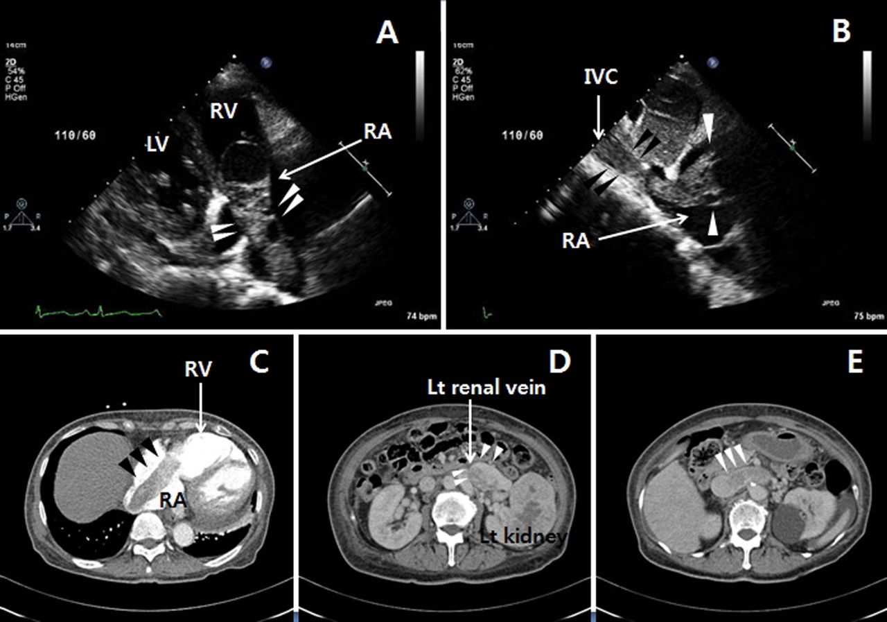
A 69-year-old woman with hypertension, with dyspnoea for 1 month, was admitted to the emergency department. On presentation, transthoracic echocardiography showed preserved left ventricular systolic function (ejection fraction=52%) and a large, highly mobile linear cystic mass in the right atrium coming from the inferior vena cava (figure 1A,B, arrowheads; online supplementary videos 1–3). Severe tricuspid stenosis was shown during the diastolic phase owing to a large mobile mass in the right ventricular inflow portion (Vmax=1.6 m/s, peak/mean diastolic pressure gradient=10/6 mm Hg).
Echocardiography ((A, B) right ventricular inflow view and subcostal view, arrowheads) and CT scan ((C–E) arrowheads) showing the huge, linear cystic mass in the right atrium (RA) coming from the inferior vena cava (IVC). LV, left ventricle; RV, right ventricle.
Abdominal CT showed a heterogeneous large mass in the left kidney and heterogeneous internal low density in the left renal vein, inferior vena cava and right atrium, suggestive of renal cell carcinoma and tumour thrombosis (figure 1C–E, arrowheads). She refused surgery and is being treated with chemotherapy for the renal cell carcinoma and anticoagulation for thrombi in the heart.
Supplementary materials
Supplementary Data
This web only file has been produced by the BMJ Publishing Group from an electronic file supplied by the author(s) and has not been edited for content.
Files in this Data Supplement:
- Data supplement 1 - Online video 1
- Data supplement 2 - Online video 2
- Data supplement 3 - Online video 3
Footnotes
-
Contributors S-JY is the first author who treated the patient and organised the examination. DWJ provided comments on this case and carried out the figure imaging; JYY treated the patient and provided comments on this case.
-
Competing interests None.
-
Patient consent Obtained.
-
Provenance and peer review Not commissioned; externally peer reviewed.