Article Text

Statistics from Altmetric.com
- Percutaneous coronary intervention (PCI)
- Intravascular ultrasound (IVUS)
- Plain old balloon angioplasty (POBA)
- coronary angioplasty
- intravascular ultrasound
A 71-year-old man, who was treated for hypertension and dyslipidaemia, had recurrent chest pain on exertion and was transferred to our hospital. He had a previous history of inferior myocardial infarction, and the right coronary artery (RCA) had been treated with plain old balloon angioplasty (POBA) 22 years ago. Four years later, this lesion became stenosed again and was re-treated with POBA.
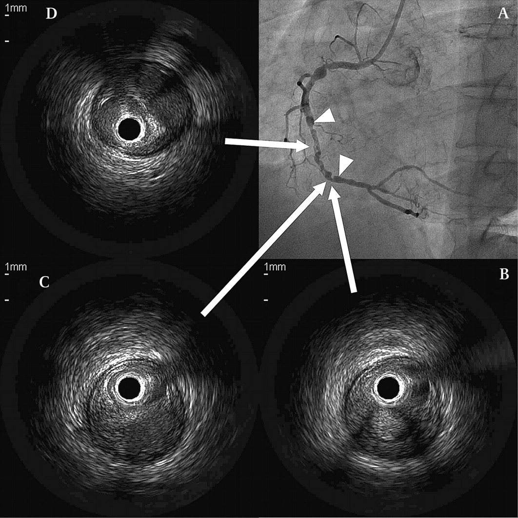
On admission, he was diagnosed as having unstable angina, and coronary angiography (CAG) was performed immediately. CAG demonstrated a severe stenosis at the left circumflex artery (LCx), and we urgently treated him with percutaneous coronary intervention (PCI). In addition to the culprit lesion of LCx, we found an unusual angiographically stenotic lesion at the site of the RCA where repeated POBA was performed previously (figure 1A and video 1).
We performed staged PCI for the RCA lesion. Pre-PCI intravascular ultrasound (IVUS) showed a spiral barrel-like appearance at the previous POBA site (figure 1B–D and video 2, figure 1B–D: each figure showed a barrel-like appearance of each point. Video 2: IVUS finding of RCA from distal arrow to proximal arrow was recorded). This finding resembled the lotus root appearance seen in Kawasaki disease.1 The RCA lesion was dilated with POBA, and then the entire lesion was covered with two drug-eluting stents. His recovery was uneventful.
The RCA lesion was thought to have been dissected following the previous repeated POBA, temporarily occluded with thrombus, then spontaneously recanalised and finally remodelled to the current appearance after 18 years.
Reference
Footnotes
Competing interests None.
Patient consent Obtained.
Provenance and peer review Not commissioned; externally peer reviewed.